Thermal Imaging
A Heat-Seeking Start to Wellness
Inflammation affects skin temperature, which makes thermal imaging an incredible screening tool and one that is especially useful in the quick detection of conditions such as arthritis, repetitive strain injury, muscular pain and circulatory problems.
After infrared cameras capture and record temperature variations on the skin, a close analysis of the images gives an accurate picture of what is going on inside the body, sometimes with surprising results.
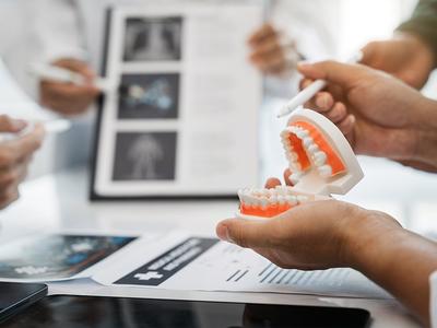
“We recently saw on our thermal imaging camera how a guest’s problem with a tooth was creating inflammation in the whole body down to the breast,” said Tina Christoudias-Spyrou, Neomed’s Functional Medicine Practitioner and Nutritionist.
How Thermal Imaging Works
Medical thermal imaging measures infrared radiation, which is then translated into thermal images and corresponding temperature calculations.
It’s a two-part process with cameras first capturing and measuring infrared radiation emitted by a patient. Via connection to a laptop or other monitor, pattern-recognition software then reveals any increases or decreases in skin surface temperature by means of a ‘heat map’.
This screening process is pain-free, non-invasive, radiation-free and an easy-to-use method to examine and monitor patients quickly and accurately.
Thermal imaging is most commonly used in the fields of rheumatology, neurology, oncology, physiotherapy and sports medicine. However, with the rise of ever-sophisticated technology, it is fast becoming the first call in the diagnosis of a wide range of illnesses and conditions.
Inflammation Markers
Thermal imaging is especially effective in diagnosing arthritis because arthritic joints usually have higher temperatures than non-arthritic joints, so thermal imaging has become a valuable tool in evaluating and monitoring inflammation caused by the early stages of the disease.
In a study conducted by Roope Lasanen from the University of Eastern Finland, thermal cameras diagnosed inflammation in knee and ankle joints in children. It was also found that skin surface temperatures were significantly elevated in inflamed ankle joints.
Elsewhere, thermal imaging has also proved effective in the diagnosis of muscular sports injuries.
A study last year into the ability of infrared thermal imaging to identify soft tissue temperature alterations as a result of exercise and as a result of injury found that thermal imaging is sensitive enough to “delineate variability in temperature that is indicative of both pathology as well as tissue strain”.
“Although exercise and injury may manifest in a similar thermal pattern, an ankle sprain manifests as a more concentrated focal heat patch covering a wider range of tissue in which thermal segmentation extends widely in the surrounding tissue,” the report concluded, adding that the screening technique was under-developed in the field of sports medicine.
Warm surprises
In some respects, the potential of thermal imaging has yet to be fully realised, but academic research continues to show results in a variety of conditions.
For example, in 2018, researchers at the University of Nottingham’s Institute for Aerospace Technology (IAT), together with academic staff from the Bioengineering and Human Factors Research Groups, found that facial temperatures were strongly correlated to mental workload.
The most affected area was above the sinuses, around the nose, with temperatures reducing as participants carried out tasks of increasing difficulty.
“The results show that when people are fully focused on a task, their breathing rate changes as the autonomic nervous system takes over. There may also be a diversion of blood flow from the face to the cerebral cortex as the mental demand increases, although this is the subject of further research.”
In the same year, researchers at the Institute of Microbiology and Hygiene at the Charité University of Medicine in Berlin found that thermal imaging flagged up a skin infestation called tungiasis, which causes inflammation of the skin.
Results showed that the temperature around a lesion caused by tungiasis was significantly higher than the median temperature of the foot. The study concluded that the technique could be used to diagnose hidden and atypical manifestations of tungiasis and other infectious skin diseases prevalent in the tropics.
As well as providing insight into mental and physical conditions, some researchers believe thermography can determine an individual’s emotional state. At the University of Granada, researchers found that when an individual viewed an image of a loved one, specific regions around the body increased in temperature by up to two degrees centigrade. Even more, interestingly, the thermographic system revealed that while passion increases temperatures around the hands and face, empathy decreases temperatures, especially in the nose.
The Advantages of Thermal Imaging
Thermal imaging can be used as an initial tool to assess overall health and for the early screening of diseases. Compared with traditional detection methods such as X-rays, ultrasounds, CT scans and endoscopic examinations, thermal imaging comes with no radiation damage, it’s non-invasive and it is pain-free. It can also be very useful when tracking the progress of treatment.
Neomed’s resident physiotherapist Lubka Mlčúchová said, “Thermal imaging or thermography is a non-invasive way to detect the areas or patterns of the body showing increased blood flow in body tissues.
“We use the infrared camera to capture heat waves emitted by the body. These are then interpreted by software that creates the thermal images.
“It's a good tool for the initial and final assessment of patients, allowing us to see the difference before and after a course of treatment.”
For more information about our Thermal Imaging Session, please fill in the form below: